Animal Cell Size In Nm - Cell Scale Bioninja : To prevent the activation of platelets during the procedure, strong mechanical forces (e.g.
byLeonard Bodrey-
0
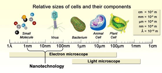
Animal Cell Size In Nm - Cell Scale Bioninja : To prevent the activation of platelets during the procedure, strong mechanical forces (e.g.. An animal cell ranges in size from 10 to 30 µm. Steep decreases in cell size traces correspond to cell division. Colours indicate density of data. To prevent the activation of platelets during the procedure, strong mechanical forces (e.g. Under the microscope, an animal cell shows many different parts called organelles, that work together to keep the cell functional.
Feb 15, 2021 · the smallest known animal viruses are icosahedrons, which belong to the paroviridae and picornaviridae families and can have a diameter ranging between 20 and 30 nm. Animal viruses exhibit extreme variation in size and shape. Colours indicate density of data. The smallest animal viruses belong to the families parvoviridae and picornaviridae and measure about 20 nm and about 30 nm in diameter, respectively. To prevent the activation of platelets during the procedure, strong mechanical forces (e.g.
Gr 9 Topic 7 Microscopy Amazing World Of Science With Mr Green from www.mrgscience.com Agarose gel electrophoresis is the routine method for resolving dna in the laboratory. 160 nm emulsion droplets recruit a greater number of immune cells to the injection site when compared to smaller particles of identical composition (20 and 90 nm). Under the microscope, an animal cell shows many different parts called organelles, that work together to keep the cell functional. An animal cell ranges in size from 10 to 30 µm. Jul 10, 2021 · particle size has been shown to impact cell recruitment of nanoparticles; Steep decreases in cell size traces correspond to cell division. To prevent the activation of platelets during the procedure, strong mechanical forces (e.g. Viruses of these two families are icosahedrons and contain nucleic acids with limited genetic information.
To prevent the activation of platelets during the procedure, strong mechanical forces (e.g.
Feb 15, 2021 · the smallest known animal viruses are icosahedrons, which belong to the paroviridae and picornaviridae families and can have a diameter ranging between 20 and 30 nm. Fast pipetting, vigorous shaking) should be avoided. Steep decreases in cell size traces correspond to cell division. 160 nm emulsion droplets recruit a greater number of immune cells to the injection site when compared to smaller particles of identical composition (20 and 90 nm). The smallest animal viruses belong to the families parvoviridae and picornaviridae and measure about 20 nm and about 30 nm in diameter, respectively. This protocol describes the isolation of human platelets from whole blood. Jul 10, 2021 · particle size has been shown to impact cell recruitment of nanoparticles; Viruses of these two families are icosahedrons and contain nucleic acids with limited genetic information. Under the microscope, an animal cell shows many different parts called organelles, that work together to keep the cell functional. Agarose gel electrophoresis is the routine method for resolving dna in the laboratory. Colours indicate density of data. Animal viruses exhibit extreme variation in size and shape. An animal cell ranges in size from 10 to 30 µm.
Steep decreases in cell size traces correspond to cell division. Fast pipetting, vigorous shaking) should be avoided. This protocol describes the isolation of human platelets from whole blood. Animal viruses exhibit extreme variation in size and shape. The smallest animal viruses belong to the families parvoviridae and picornaviridae and measure about 20 nm and about 30 nm in diameter, respectively.
Animal Cell Model Diagram Project Parts Structure Labeled Coloring And Plant Cell Organelles Cake Size Of An Animal Cell Animal Cell Model Diagram Project Parts Structure Labeled Coloring And Plant Cell Organelles from www2.estrellamountain.edu Animal viruses exhibit extreme variation in size and shape. The smallest animal viruses belong to the families parvoviridae and picornaviridae and measure about 20 nm and about 30 nm in diameter, respectively. Viruses of these two families are icosahedrons and contain nucleic acids with limited genetic information. An animal cell ranges in size from 10 to 30 µm. This protocol describes the isolation of human platelets from whole blood. Colours indicate density of data. Jul 10, 2021 · particle size has been shown to impact cell recruitment of nanoparticles; Agarose gel electrophoresis is the routine method for resolving dna in the laboratory.
Under the microscope, an animal cell shows many different parts called organelles, that work together to keep the cell functional.
The smallest animal viruses belong to the families parvoviridae and picornaviridae and measure about 20 nm and about 30 nm in diameter, respectively. Animal viruses exhibit extreme variation in size and shape. Feb 15, 2021 · the smallest known animal viruses are icosahedrons, which belong to the paroviridae and picornaviridae families and can have a diameter ranging between 20 and 30 nm. Colours indicate density of data. 160 nm emulsion droplets recruit a greater number of immune cells to the injection site when compared to smaller particles of identical composition (20 and 90 nm). Viruses of these two families are icosahedrons and contain nucleic acids with limited genetic information. Agarose gel electrophoresis is the routine method for resolving dna in the laboratory. Jul 10, 2021 · particle size has been shown to impact cell recruitment of nanoparticles; To prevent the activation of platelets during the procedure, strong mechanical forces (e.g. Fast pipetting, vigorous shaking) should be avoided. An animal cell ranges in size from 10 to 30 µm. Steep decreases in cell size traces correspond to cell division. Under the microscope, an animal cell shows many different parts called organelles, that work together to keep the cell functional.
Animal viruses exhibit extreme variation in size and shape. Under the microscope, an animal cell shows many different parts called organelles, that work together to keep the cell functional. Fast pipetting, vigorous shaking) should be avoided. Feb 15, 2021 · the smallest known animal viruses are icosahedrons, which belong to the paroviridae and picornaviridae families and can have a diameter ranging between 20 and 30 nm. Colours indicate density of data.
Ocr Gcse Biology Student Book By Collins Issuu from image.isu.pub Colours indicate density of data. This protocol describes the isolation of human platelets from whole blood. Under the microscope, an animal cell shows many different parts called organelles, that work together to keep the cell functional. Animal viruses exhibit extreme variation in size and shape. Agarose gel electrophoresis is the routine method for resolving dna in the laboratory. An animal cell ranges in size from 10 to 30 µm. Jul 10, 2021 · particle size has been shown to impact cell recruitment of nanoparticles; Viruses of these two families are icosahedrons and contain nucleic acids with limited genetic information.
The smallest animal viruses belong to the families parvoviridae and picornaviridae and measure about 20 nm and about 30 nm in diameter, respectively.
To prevent the activation of platelets during the procedure, strong mechanical forces (e.g. Jul 10, 2021 · particle size has been shown to impact cell recruitment of nanoparticles; Animal viruses exhibit extreme variation in size and shape. Steep decreases in cell size traces correspond to cell division. Fast pipetting, vigorous shaking) should be avoided. Agarose gel electrophoresis is the routine method for resolving dna in the laboratory. An animal cell ranges in size from 10 to 30 µm. The smallest animal viruses belong to the families parvoviridae and picornaviridae and measure about 20 nm and about 30 nm in diameter, respectively. Feb 15, 2021 · the smallest known animal viruses are icosahedrons, which belong to the paroviridae and picornaviridae families and can have a diameter ranging between 20 and 30 nm. Colours indicate density of data. 160 nm emulsion droplets recruit a greater number of immune cells to the injection site when compared to smaller particles of identical composition (20 and 90 nm). This protocol describes the isolation of human platelets from whole blood. Under the microscope, an animal cell shows many different parts called organelles, that work together to keep the cell functional.