Plant Cell Under Microscope Drawing / Cell Structure : Major differences between a plant cell and on animal cell are (i) presence of chloroplast in plant cell.
byLeonard Bodrey-
0
Plant Cell Under Microscope Drawing / Cell Structure : Major differences between a plant cell and on animal cell are (i) presence of chloroplast in plant cell.. Cell is the basic building blocks of all organisms. Living cells, however, can be observed directly under phase contrast for measuring the actual size of a cell or a microscopic plant organ a stage micrometer and an ocular scale or micrometer are necessary. Microscope slide cover slip onion. Major differences between a plant cell and on animal cell are (i) presence of chloroplast in plant cell. Use them in commercial designs under lifetime, perpetual & worldwide rights.
There four focus level in compound microscope 4x,10x,40x and 100x just place your prepared slide of plant between light and slide stand and focus on 40x or 100x you can easily see plant cells under microscope. The ocular lens (or eye piece) which magnifies an object there are three structures that distinguish plant cells from animal cells. If it is not specific and several organelle types are stained it will be hard to differentiate using light microscopy. Make it about 10 cm long. The high resolving power makes the electron microscope a very important research tool in microbiology.
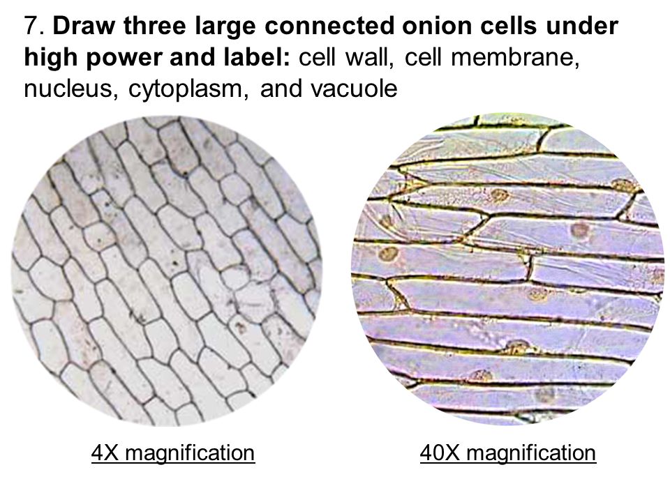
Lab Comparing Plant And Animal Cells Ppt Video Online Download from slideplayer.com Observe the onion skin under low power of the microscope and then under high power. Draw a single cell as seen under the higher power. Cells consist of cytoplasm enclosed within a membrane, which contains many biomolecules such as proteins and nucleic acids.2 most plant and animal cells are only visible under a light microscope, with dimensions between 1 and 100 micrometres.3 electron microscopy gives a much higher. Published on december 9, 2013 at 4:03pm by glenda stovall under cell. Label these structures in your high. Ever since the first microscope was used, biologists have been ch lab # objective: Use them in commercial designs under lifetime, perpetual & worldwide rights. This should result in a more stimulating experience, as you use the microscope to discover the microscopic structure of plant cells and their interrelationships with each other.
Cells are microscopic and can only be seen under a microscope.
Select the lowest power objective lens. (iii) presence of cell wall. To look at a cell close up we need a microscope. Cell structure i nucleus medical media. Animal cells introduction background information: Plant and animal cells have cell membranes, cytoplasm, a nucleus and organelles such as mitochondria and sometimes vacuoles. Find the perfect plant cells under microscope stock photos and editorial news pictures from getty images. Published on december 9, 2013 at 4:03pm by glenda stovall under cell. Examining plant cells under the microscope. So, the cells which are observed under light microscope are, actually, dead cells. Plant cells under a microscope. Structure of a plant cell. Dreamstime is the world`s largest stock photography community.
Animal cells introduction background information: Probably the only organelle you might pick out without staining or marking in some way might be the nucleus (if you. This image reminds me of something you would look at under a microscope such as cells or organisms. Image:plant cell seen under electron microscope. Structure of a plant cell.
Lab Comparing Plant And Animal Cells Ppt Video Online Download from slideplayer.com This should result in a more stimulating experience, as you use the microscope to discover the microscopic structure of plant cells and their interrelationships with each other. Plant cells under a microscope. Select the lowest power objective lens. The ocular lens (or eye piece) which magnifies an object there are three structures that distinguish plant cells from animal cells. Cell structure i nucleus medical media. The differences between plant and animal cells. (ii) presence of large central vacuole in plant cell. Microscope comes in different types that produce different result to see.
Published on december 9, 2013 at 4:03pm by glenda stovall under cell.
Microscope slide cover slip onion. See more ideas about microscopic photography, cell drawing, patterns in nature. It is such a simple image yet it seems to hold a lot of detail. Dreamstime is the world`s largest stock photography community. They are very tiny than what human eyes can see in general. Make it about 10 cm long. Use them in commercial designs under lifetime, perpetual & worldwide rights. Cells are microscopic and can only be seen under a microscope. Given below is the diagram of a cell as seen under the microscope after having been placed in a solution Lets get looking at some real plant cells! The cell membrane encloses the contents of the cell and separates it from its environment. Image:plant cell seen under electron microscope. Plant and animal cells have cell membranes, cytoplasm, a nucleus and organelles such as mitochondria and sometimes vacuoles.
The high resolving power makes the electron microscope a very important research tool in microbiology. Label these structures in your high. Ever since the first microscope was used, biologists have been ch lab # objective: They are very tiny than what human eyes can see in general. Plant and animal cells have cell membranes, cytoplasm, a nucleus and organelles such as mitochondria and sometimes vacuoles.
Illustrate Only A Plant Cell As Seen Under Electron Microscope How Is It Different From Animal Cell Sarthaks Econnect Largest Online Education Community from www.sarthaks.com They are very tiny than what human eyes can see in general. Browse 153 plant cells under microscope stock photos and images available, or start a new search to explore more stock photos and images. 8 pictures of plant cells under a microscope. (ii) presence of large central vacuole in plant cell. (refer to box 7.1 on p. This should result in a more stimulating experience, as you use the microscope to discover the microscopic structure of plant cells and their interrelationships with each other. Answer the following questions in your exercise book. A cell is a very tiny structure which exists in living bodies.
Answer the following questions in your exercise book.
(iii) presence of cell wall. Cell is the basic building blocks of all organisms. The ocular lens (or eye piece) which magnifies an object there are three structures that distinguish plant cells from animal cells. To learn how to get the best image from a microscope. Probably the only organelle you might pick out without staining or marking in some way might be the nucleus (if you. Answer the following questions in your exercise book. The cell membrane encloses the contents of the cell and separates it from its environment. Animal cells introduction background information: The differences between plant and animal cells. 8 pictures of plant cells under a microscope. A cell is a very tiny structure which exists in living bodies. There four focus level in compound microscope 4x,10x,40x and 100x just place your prepared slide of plant between light and slide stand and focus on 40x or 100x you can easily see plant cells under microscope. Plant cells under the microscope.
Browse 153 plant cells under microscope stock photos and images available, or start a new search to explore more stock photos and images plant cell microscope drawing. To learn how to get the best image from a microscope.